Article Text

Abstract
Background: Noise exposure in neonatal units has long been suspected of being a cause of hearing loss associated with such units. The noise intensity to which the neonate is exposed varies with the type of ventilatory support used. Also, the post-nasal space is an enclosed cavity that is close to the inner ear and an area of turbulent and hence potentially noisy airflow.
Aim: To determine noise intensities within the ear and post-nasal space in neonates on different modes of ventilatory support using probe microphones, measures previously not undertaken.
Methods: A portable instrument with a probe microphone was used for the measurements. Three groups of infants were included: (a) those receiving no respiratory support (NS); (b) those receiving conventional ventilation (CV); (c) those receiving continuous positive airways pressure (CPAP) support.
Results: The mean in-the-ear noise intensities (at 1 kHz) were 41.7 dB SPL (NS), 39.5 dB SPL (CV), and 55.1 dB SPL (CPAP). The noise intensities in the post-nasal space in those receiving CPAP support were higher than in the other groups, reached mean levels of up to 102 dB SPL at some frequencies, and increased with increasing flow rates.
Conclusions: The most important finding is the high noise intensities in the post-nasal space of those receiving CPAP support. Given the proximity of the post-nasal space to the inner ear, enough noise could be transmitted, especially in infants receiving the higher flow rates, to cause cochlear damage and hence hearing loss. It would therefore be wise, wherever possible, to avoid using the higher flow rates.
- hearing loss
- deafness
- cochlear damage
- noise intensities
- intensive care units
Statistics from Altmetric.com
Neonates admitted to a neonatal intensive care unit are 10.2 times more likely to have a sensorineural or mixed hearing loss than those who are not.1 Of all the cases of sensorineural hearing loss in children, 12.7% have been ascribed to perinatal factors.2 Borg3 in a detailed review of the literature concluded that permanent damage of the inner ear could result from hypoxia especially if it is recurrent or prolonged and particularly if it occurs in combination with ischaemia. However, he found that the best predictors for sensorineural hearing loss of perinatal origin are the duration of artificial ventilation and length of stay on the neonatal unit. Noise exposure in neonatal intensive care units, either acting alone or more likely in synergy with other adverse factors such as hypoxia and ototoxic drugs,4,5 has long been suspected of being a causative factor in sensorineural hearing loss associated with neonatal intensive care units. Indeed, animal studies have shown that the young cochlea, whether premature or full term, is more susceptible to the damaging effects of noise than the mature adult organ.6–8 Outer hair cell damage has been shown in young guinea pigs exposed to noise levels up to 80 dB SPL.7
Noise is also a potential cause of stress in the neonatal unit,9,10 the threshold for stimulation of the adrenopituitary axis being 68 dB linear.11 Levels of noise as low as 70 dB SPL12 have produced effects on the cardiovascular system, such as peripheral vasoconstriction, increased heart rate, and raised blood pressure. Noise levels of 50–75 dBA have been shown to produce significant sleep disturbance in infants.13 Zahr and Traversay14 found that the use of ear muffs led not only to significantly higher oxygen saturation levels and fewer behavioural changes (indicators of lower stress levels), but also resulted in neonates sleeping for longer.
Although numerous studies have been performed to determine noise levels on neonatal units, they have been carried out using sound level meters7,14–16 rather than probe microphones. Further, no study has measured noise levels in the post-nasal space, a region of potentially turbulent and hence noisy airflow, which is close to the middle and inner ear, particularly in neonates. The use of the probe microphone allows measurement of noise intensities within the post-nasal space as well as within the ear canal, something that cannot be done with sound level meters.
The aim of this study was to measure the levels of noise in the ear and post-nasal space, using probe microphones, of three groups of neonates: (a) those receiving no respiratory support, (b) those receiving conventional ventilation, and (c) those receiving continuous positive airways pressure (CPAP) support.
MATERIALS AND METHODS
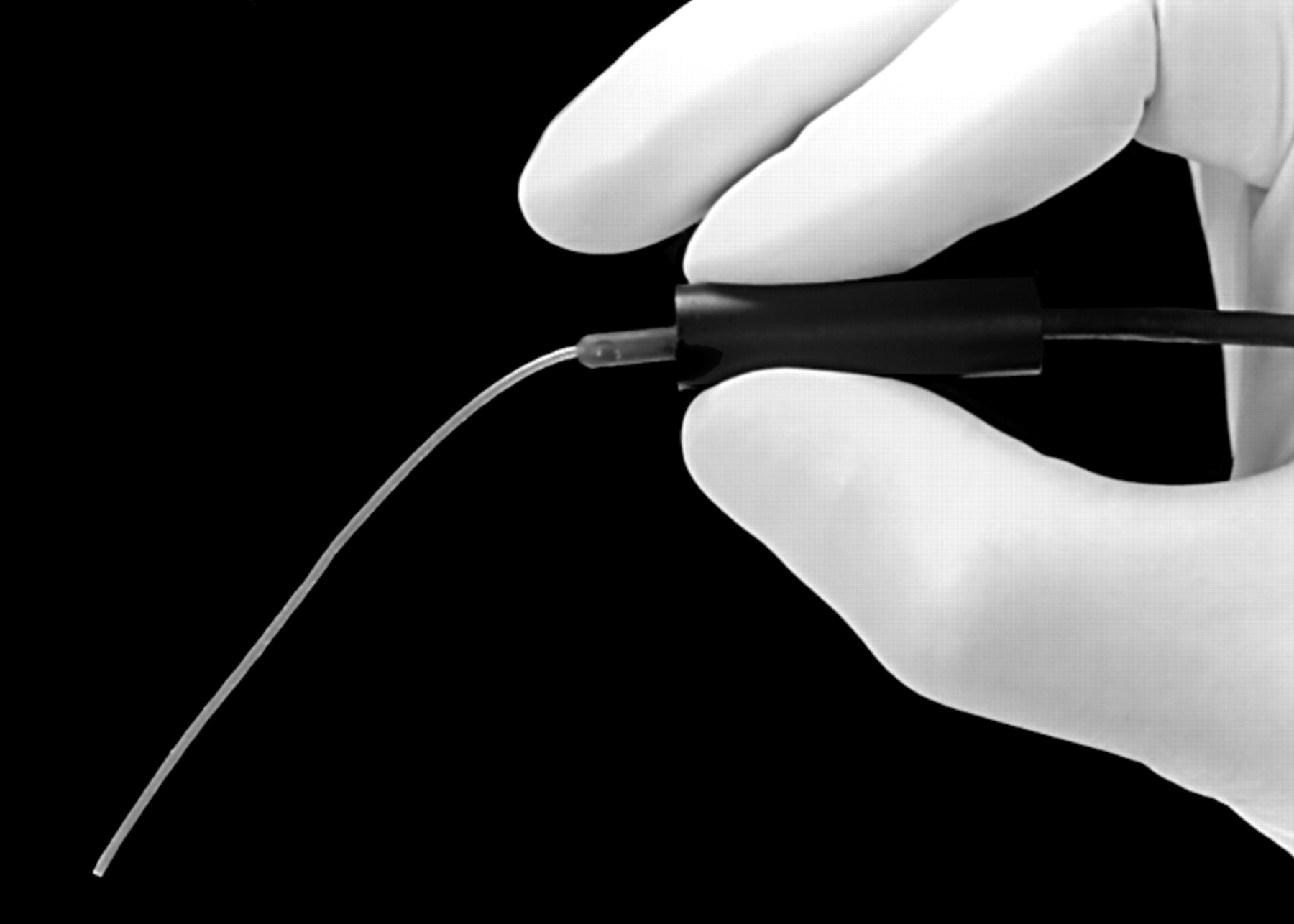
Subjects for the study were selected from preterm neonates admitted to a neonatal intensive care unit. Only those deemed medically stable and who had no aural or nasal malformations were recruited. They also had to have been on the unit for at least 24 hours before being considered for inclusion. This was to ensure that the baby was clinically stabilised and that the parents had had a chance to settle before being approached about the study. Three groups of patients, all of whom were nursed in incubators, were recruited: (a) those not having any respiratory support; (b) those on conventional ventilation (all were on the SLE 2000 ventilator); (c) those on CPAP support (all were nasal prong via Infant Flow Driver). During the period of the study, only two patients were placed on high frequency ventilation, but both were too unwell to be included in the study. Measurements were carried out using a probe microphone/tube (fig 1) and a Fonix FP40 analyser, which is capable of measuring noise intensities at 0.2, 0.5, 1, 2, 4, and 8 kHz. All measurements were made at the same time of day (0700–0730) to minimise variability caused by extraneous noise.

Probe microphone/tube used to measure noise intensity.
Terminology
The range of sound pressures over which humans can hear is enormous; the largest sound pressure (loudest noise) that can be tolerated is 10 million times greater than the smallest (quietest noise) that can be sensed. For practical purposes, dealing with such a range of values is too cumbersome. To overcome this difficulty, the logarithmic decibel (dB) scale was introduced; on this scale the values are reduced to a more manageable 0 and 140. To be meaningfully measured, a sound has to be compared with a reference value. The reference level most commonly used is the quietest sound pressure level that can be heard (20 μPa) and thus the sound is expressed as dB SPL (sound pressure level). Although the sound is measured in dB SPL with a sound level meter, different “weightings” can be used. In dBA measurements, the low frequencies are attenuated, just as the human auditory system does. In dB linear measurements, all the frequencies are given equal “weight”.
The probe tube is of narrow calibre and easily blocked by debris and mucus, which could result in falsely low measurements of noise intensity in the ear canal and post-nasal space. For this reason, two measurements were made for each ear: one at the opening of the ear canal and one within the external auditory meatus. Similarly, noise levels in the post-nasal space were determined first just outside the nasal opening and then by sliding the probe tube through the anterior nares, along the floor of the nasal cavity, and into the post-nasal space. The external measurement in each case served as a reference to indicate whether measurements in the ear canal and post-nasal space were realistic representations of the noise intensities in these sites. If the latter were substantially lower than the respective external reference measurements, it was highly likely that the probe tube had become blocked. In such cases, it was withdrawn and visually inspected. If it was blocked, it was replaced with a new one. If it was deemed patent, it was reinserted and the measurements repeated. If there were no discrepancies as just outlined, then a single measurement was obtained for each defined location. All the measurements were obtained by the same researcher.
The t test for independent samples was used to determine whether or not there was any significant difference between:
(a) noise levels outside and within the ear canal for each frequency and for each group of subjects;
(b) noise levels within the ear of those receiving no respiratory support and ventilated subjects;
(c) noise levels within the ear of ventilated patients and those on CPAP;
(d) noise levels within the ear of those with no respiratory support and those on CPAP;
(e) noise levels in the post-nasal space of those with no respiratory support and ventilated subjects;
(f) noise levels in the post-nasal space of ventilated subjects and those on CPAP.
RESULTS
Twenty two subjects were recruited to the study as follows: (a) five neonates on no respiratory support; (b) five neonates on conventional ventilation (SLE 2000 ventilator); (c) 12 neonates on CPAP support (nasal prong via Infant Flow Driver). The gestation range was 27–32 weeks.
Table 1 shows the mean noise levels for these three groups and gives the intensity levels for outside the ear (Out) and within the external canal (In). Comparison of these for each frequency, using the t test for independent samples, shows no significant difference between the two sets of means for each group.
Mean noise intensities in dB SPL (in and out of ears)
Table 2 shows the results of statistical comparison between the mean noise levels in the ears for those on no respiratory support and those on conventional ventilation. It can be seen that there is no significant difference between the two groups. Table 2 also shows the results of comparison between those being ventilated and those on CPAP support and also a comparison between those on no ventilatory support and those on CPAP. It can be seen that, for frequencies 0.5–8 kHz, those on CPAP were exposed to significantly higher noise intensities than either those receiving no respiratory support or those being ventilated. For 0.2 kHz there was no significant difference in either comparison.
Comparison of mean in the ear noise intensities between groups
Comparison between the mean noise intensity levels, across the frequency range, outside the nose and within the post-nasal space for those on no respiratory support and those on conventional ventilation showed no significant difference. As the measurements in the post-nasal space were made at different flow rates for those on CPAP support, such a statistical comparison was not possible, but all outside measurements were found to be generally lower than the post-nasal ones, even at the lowest flow rates.
Table 3 shows that the mean noise intensities in the post-nasal space of ventilated neonates were significantly higher at 0.5, 1, and 2 kHz than in those on no respiratory support. Table 3 also shows that mean noise intensities in the post-nasal space in the neonates on CPAP at the lowest flow rate (5 litres/min) was significantly higher across the frequency range than for those on conventional ventilation.
Comparison of post-nasal space noise levels
Table 4 shows the mean noise intensities in the post-nasal space for different flow rates. Figure 2 is a graphical representation for three of the flow rates (5, 8, and 10 litres/min). It can be seen that mean noise intensity generally increases across the frequency range with increase in flow rate. Intensity levels are particularly high for flow rates of 9 and 10 litres/min.
Mean noise levels in the post-nasal space for different flow rates of CPAP

Graphical representation of increase in post-nasal noise intensity for selected flow rates.
DISCUSSION
In previous studies,7,14,16,17 noise intensities on neonatal units were determined using sound level meters measuring noise levels in dBA and are therefore not directly comparable with the measurements made in this study, which were in sound pressure levels (SPL) at discrete frequencies. However, the same reference measure is used in calibrating both the sound level meter and the Fonix FP40 analyser, and therefore the noise intensities at 1 kHz are comparable.18,19 The mean ear noise intensity found in this study (table 1) for the three groups of subjects at 1 kHz is considerably less than that found in other studies.7,14,16,17 Neonates on conventional ventilation are not exposed to significantly more noise than those who have no respiratory support. It should be noted, however, that, although not statistically significant, the real ear values for those on conventional ventilation are actually less, across the frequency range, than for those on no respiratory support. A possible explanation is that the natural breathing of the neonates with no respiratory support creates turbulence in the upper respiratory passages and is thus actually noisier than the mechanical ventilation of those on a modern ventilator. In ventilated patients, the airflow and any turbulence generated is downwards, through an endotracheal tube, into the trachea and away from the post-nasal space and ears. Another possible factor is the quality of modern ventilatory equipment. Babies on CPAP support are exposed to significantly more noise than the other two groups. The noise intensities lie in the range 53–57 dB SPL (55 dB SPL at 1 kHz), a level considerably above the upper limit of 45 dBA recommended by the Committee on Environmental Health of the American Academy of Pediatrics.20
Kruger and Ruben21 showed that the resonance frequency of the external ear canal is a function of the volume of the external canal and varies with age, being on average 7.2 kHz for newborns and 2.7 kHz for adults. It is likely that, for the preterm subjects in this study, the resonance frequency of the external auditory meatus was very much greater than the 7.2 kHz recorded for term newborns and hence beyond the maximum 8 kHz measured in this study. This explains why no significant difference was observed between noise levels outside and within the external canals in all three groups of neonates.
Noise intensities in the post-nasal space are significantly higher at 0.5, 1, and 2 kHz for those on conventional ventilation than those on no respiratory support (table 3). Even so, these intensity levels are not excessive in terms of the amount of noise that may be transmitted to the inner ear by means of bone conduction. By contrast, noise levels in the post-nasal space of the babies on CPAP support at one of the lowest flow rates used clinically (5 litres/min) are significantly greater across the frequency range than for those on conventional ventilation (table 3). For the purposes of pure tone audiometry, it is considered that there is a transcranial attenuation of air conducted sound of 40 dB SPL for adults. Bearing in mind that the distance and intervening soft tissue between the post-nasal space and the inner ear in neonates is far less, it is likely that at least 40 dB SPL on average is being transmitted to the inner ear in neonates on minimal flow rates (table 3). Another means by which noise can reach the ears of neonates is via the Eustachian tube. This mode of transmission would be further facilitated by the fact that CPAP produces a positive pressure encouraging the opening of the Eustachian tube. As the flow rates are increased, noise intensities in the post-nasal space also increase (table 4). For example, at a flow rate of 10 litres/min, a mean noise level of 100.3 dB SPL at 1 kHz is generated in the post-nasal space. Therefore at least 60 dB SPL of noise is theoretically being transmitted to the inner ear of these neonates. It should be borne in mind that this is only the mean, and that levels of up to 115 dB SPL were obtained in some cases at the higher flow rates, giving a theoretical transmission of at least 75 dB SPL to the inner ear. It should be noted that the findings in this study relate to the devices used—that is, SLE 2000 ventilator and nasal prongs with an Infant Flow Driver—and cannot be extrapolated to other devices.
There are several unknown factors that prevent any firm conclusions being reached about cochlear damage in neonates. Firstly, although animal experiments have indicated that the immature cochlea is more susceptible to noise induced hearing loss than the adult organ,7 the exact situation in the human neonate is not known. Secondly, although it is widely thought, and has been shown in animal experiments, that synergism of noise with other adverse factors such as aminoglycoside antibiotics is a likely mechanism of the hearing loss,3–5 the situation in humans is not entirely clear. Thirdly, it has been shown, again in animal models, that there is a period after birth when the immature cochlea is especially sensitive to acoustic trauma.22 Whether this is also true for the human infant, particularly the preterm baby, is far from certain. Finally, it is difficult to be sure, without any means of accurate measurement, exactly how much noise is being transmitted from the post-nasal space of neonates on CPAP to the inner ear. Given these uncertain factors, further investigations into the possible role of CPAP, particularly in infants nursed for long periods of time on it, in sensorineural hearing loss associated with adverse perinatal factors and stay in a neonatal intensive care unit seem warranted.