Article Text

Abstract
Objective We aimed to identify gestational-age corrected prenatal ultrasound markers of complex gastroschisis, and to compare physical growth and neurodevelopment between children with simple and complex gastroschisis.
Design We included prenatally diagnosed gastroschisis patients from 2000 to 2012 who joined our longitudinal follow-up programme. Associations between complex gastroschisis and prenatal ultrasound markers collected at 30 weeks’ gestation and prior to delivery were tested using logistic regression. Physical growth (SD scores (SDS)), mental and psychomotor developmental index (MDI, PDI; Bayley Scales of Infant Development) were recorded at 12 and 24 months. Data were analysed using general linear models and compared with population norms.
Results Data of 61 children were analysed (82% of eligible cases). Extra-abdominal bowel dilatation at 30 weeks’ gestation was significantly associated with complex gastroschisis (OR (95% CI): 5.00 (1.09 to 22.98)), with a high negative (88%) but low positive (40%) predictive value. The mean (95% CI) height SDS at 12 months (−0.46 (–0.82 to –0.11)), and weight SDS at 12 and 24 months (−0.45 (–0.85 to –0.05), and −0.44 (−0.87 to –0.01), respectively) fell significantly below 0 SDS. MDI and PDI were significantly below 100 at 24 months; 93 (88 to 99) and 83 (78 to 87), respectively). Children with complex gastroschisis had a significantly lower PDI (76 (68 to 84)) than those with simple gastroschisis (94 (90 to 97), p<0.001).
Conclusions Prenatal ultrasound markers could not reliably distinguish between simple and complex gastroschisis. Children with complex gastroschisis may be at increased risk for delayed psychomotor development; they should be monitored more closely, and offered timely intervention.
- gastroschisis
- abdominal wall defect
- outcome
- follow-up
- growth
- neurodevelopment
Statistics from Altmetric.com
What is already known on this topic?
Various thresholds of bowel dilatation are used for predicting complex gastroschisis.
Neurodevelopmental outcome in gastroschisis patients is favourable; however, no previous study differentiated between simple and complex gastroschisis.
What this study adds?
Extra-abdominal bowel dilatation at 30 weeks’ gestation may indicate complex gastroschisis, although with low positive predictive value.
Children with complex gastroschisis may be at risk for delayed psychomotor development.
Introduction
Gastroschisis is a congenital abdominal wall defect with an estimated prevalence of 2.16 per 10 000 pregnancies.1 Surgery is required shortly after birth, by means of primary closure when possible or by placing a silastic silo to allow gradual reduction into the abdominal cavity prior to definite closure.2
Gastroschisis patients who have additional intestinal defects, that is, intestinal atresia, perforation, necrosis or volvulus (‘complex’ gastroschisis),3 have a higher risk of morbidity than children with ‘simple’ gastroschisis (without intestinal defects). This includes prolonged time to full enteral feeding (TFEF) and prolonged length of hospital stay (LOS).3–5
Over 90% of gastroschisis cases are diagnosed prenatally.6 A recent meta-analysis evaluated several prenatal ultrasound markers and showed significant positive associations between intra-abdominal bowel dilatation (IABD) and intestinal atresia, between polyhydramnios and intestinal atresia, and between gastric dilatation and neonatal death.7 These findings should be interpreted with caution, however, given that definitions of bowel and gastric dilatation differed between studies and data were not always corrected for gestational age (GA).
Additionally, adequate parental counselling should include expectations of the child’s physical growth and neurodevelopment. Physical growth in children older than 1 year has only been studied in small groups (<40 patients).8–11 No previous study has evaluated possible differences in neurodevelopment between children with simple and complex gastroschisis.
The aim of our study was to (1) identify GA-corrected prenatal ultrasound markers of complex gastroschisis and (2) to assess physical growth and neurodevelopment up to 2 years of age of children with either simple or complex gastroschisis.
Methods
Study population
A retrospective analysis was performed of prospectively collected data of all prenatally diagnosed gastroschisis cases delivered and treated at the Erasmus Medical Centre-Sophia Children’s Hospital Rotterdam between 2000 and 2012. Following the diagnosis, several prenatal characteristics were assessed every 4 weeks. From 2007 onwards, additional assessments were scheduled weekly, starting at 30 weeks’ gestation.12 Vaginal delivery was planned from 37 weeks onwards, unless obstetric reasons required otherwise. Survivors could join a longitudinal prospective follow-up programme which since 1999 is standard of care for children with anatomical congenital malformations treated in our hospital.13 The Medical Ethical Review Board waived approval (‘Medical Research in Human Subjects Act does not apply to this research proposal’; MEC-2015–308).
Prenatal and perinatal characteristics of simple and complex gastroschisis
We obtained the following ultrasound data at 30 weeks’ gestation and at the last ultrasound examination prior to delivery: amniotic fluid index, considered abnormal if <5 cm (oligohydramnios) or >24 cm (polyhydramnios); intrauterine growth restriction, defined as estimated fetal weight ≤10th percentile for GA according to the Hadlock formula III14; IABD and extra-abdominal bowel dilatation (EABD) determined using a GA-specific nomogram, considering the bowel dilated if ≥13 mm at a GA of 25–30 weeks, ≥16 mm at 30–35 weeks, and if ≥26 mm at 35–40 weeks15; and intra-abdominal gastric dilatation, defined as measurements exceeding two SDs above the mean reference value, adjusted for GA.16 Preterm birth was defined as delivery prior to 37 weeks’ gestation. Small for GA was diagnosed if birth weight was below the 10th centile according to Dutch reference curves.17 Socioeconomic status scores (with population mean 0 and SD 1) were based on postal codes.18 19
Postnatal outcome in simple and complex gastroschisis
Complex gastroschisis was defined as presence of intestinal atresia, necrosis, perforation and/or volvulus at primary postnatal evaluation. We recorded duration of initial mechanical ventilation, LOS of the initial hospitalisation, and additionally during follow-up: TFEF; number of procedures under general anaesthesia; complications; and presence of intestinal failure, defined as TFEF ≥6 weeks.20 If TFEF or LOS was ≥2 years, the duration was set at 730 days.
Physical growth and neurodevelopment
Height and weight were measured at 12 and 24 months (corrected for preterm birth). SD scores (SDS) were determined according to Dutch reference norms, with −2 to +2 SD considered as normal range.21 Neurodevelopment was assessed using the Dutch version of the Bayley Developmental Scales (BOS 2–30)22 and from December 2003, Bayley Scales of Infant Development-Second Edition (BSID-II-NL).23 These tests are interchangeable,23 and provide a mental developmental index (MDI) and psychomotor developmental index (PDI) with a mean score of 100 and an SD of 15.22 23Scores 70–84 indicate mildly impaired development, scores 55–69 moderately impaired development, and scores <55 (recorded as 55) are indicative of severe developmental delay.
Statistical analysis
Categorical variables are presented as number (%) and continuous variables as median (IQR). Prenatal characteristics and postnatal outcome parameters of children with simple or complex gastroschisis were compared using chi-square tests or Fisher’s exact tests (in case of expected counts <5) for categorical data, and the Mann-Whitney test for continuous data. We used logistic regression to find relevant ultrasound predictors of complex gastroschisis. Spearman’s rank correlation coefficient was used to assess the association between GA at birth and TFEF. General linear models were used to analyse the repeated growth and neurodevelopment measurements over time. These models included both the time point (12 or 24 months) and the type of gastroschisis (simple or complex) as independent variables. To account for the within-subject correlations, an unstructured error covariance matrix for the repeated measurements of each patient was used in the general linear models. The results are presented using estimated marginal means (ie, the predicted values of the dependent variable adjusted for covariates in the model). Estimated marginal means and their 95% CIs were compared with reference norms. A two-sided p value of <0.05 was considered statistically significant. Statistical analyses were performed using SPSS V.21.0.
Results
Of 82 prenatally diagnosed cases of gastroschisis, three (4%) pregnancies were terminated. Four (5%) other fetuses died in utero between 20 and 36 weeks’ gestation. All four showed intrauterine growth restriction but not bowel or gastric dilatation. Of 75 liveborn children, 12 (16%) were diagnosed with complex gastroschisis. One infant with simple gastroschisis died of sepsis at 5 months of age. Of 61 (82 %) children who joined the follow-up programme, 46 (75%) were seen at both 12 and 24 months (figure 1). Prenatal, perinatal and postnatal data did not significantly differ between children who joined the follow-up programme and children who did not (data not shown).

Inclusion flowchart. *Reasons for missing neurodevelopmental data at 12 months: non-cooperative n=2 (both motor/mental n=1; mental n=1); immobilisation of foot n=1 (motor n=1). At 24 months: non-cooperative n=6 (both motor/mental n=2, motor n=4); organisational n=3 (both motor/mental n=1; motor n=2), refusal n=1 (both motor/mental).
Prenatal and perinatal characteristics
Ultrasound examination at 30 weeks’ gestation revealed EABD in 6/51 (12%) fetuses postnatally diagnosed with simple gastroschisis, versus 4/10 (40%) fetuses postnatally diagnosed with complex gastroschisis (OR (95% CI) 5.00 (1.09 to 22.98)). Thus the positive and negative predictive values of EABD for complex gastroschisis were 40% and 88%, respectively. These four children with complex gastroschisis all had intestinal atresia. No significant associations were found regarding any other assessed prenatal parameter at 30 weeks’ gestation or at the last ultrasound examination prior to delivery. Perinatal data did not significantly differ between children with simple and children with complex gastroschisis (table 1).
Maternal, prenatal and perinatal characteristics of children in follow-up (n=61).
Postnatal outcome
Children with complex gastroschisis needed over three times as many procedures under general anaesthesia as those with simple gastroschisis (table 2). Moreover, median duration of initial mechanical ventilation was 24 days in infants with complex gastroschisis versus 2 days in infants with simple gastroschisis. Median durations of parenteral nutrition and hospitalisation were less than 2 months in children with simple gastroschisis, and close to 6 months in those with complex gastroschisis. There was no significant correlation between GA at birth and TFEF (Spearman’s r=−0.086, p=0.47). Intestinal failure developed in all children with complex gastroschisis, and in 25% of those with simple gastroschisis. Complications were common in both groups, especially sepsis and parenteral nutrition-related cholestasis.
Postnatal characteristics of children in follow-up (n=61).
Physical growth and neurodevelopment
Physical growth data of all gastroschisis patients in follow-up is shown in figure 2. The general linear model analysis showed significant improvement in height SDS from 12 to 24 months of +0.16 (95% CI: 0.01 to 0.31). The estimated marginal means (95% CI) of height SDS at 12 months (−0.46 (–0.82 to –0.11)), and weight SDS at 12 and 24 months (−0.45 (-0.85 to –0.05), and −0.44 (−0.87 to–0.01), respectively) fell significantly below 0 SD, but within the normal range of −2 to +2 SD. Growth parameters did not differ significantly between children with simple and complex gastroschisis. At 24 months, 4/53 (8%) children scored below −2 SD for weight; all four had simple gastroschisis. None of the children had abnormally low height SDS.
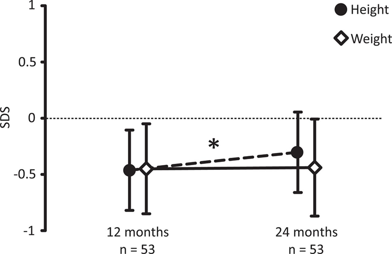
Physical growth at 12 and 24 months of all gastroschisis patients in follow-up (estimated marginal means and 95% (CI)). A = Height, B = Weight. SDS, SD score. * Indicates a significant improvement in SDS (p<0.05).
Both MDI and PDI significantly declined over time (mean differences (95% CI): −8 (−3 to –13) and −4 (−1 to –8), respectively). The estimated marginal mean (95% CI) MDI at 24 months (93 (88 to 99)) was significantly below 100, but within the normal range of 85–115. The estimated marginal mean PDI was significantly below 100 at both 12 and 24 months, 87 (82 to 92) and 83 (78 to 87), respectively. Children with complex gastroschisis scored a significantly lower PDI than those with simple gastroschisis (76 (68 to 84) vs 94 (90 to 97), respectively, p<0.001), whereas MDI did not significantly differ between groups. At 24 months a higher percentage of children with simple gastroschisis versus children with complex gastroschisis showed normal mental development (86% vs 57%, figure 3), but this difference was not statistically significant (p=0.11). The same holds for psychomotor development (81% vs 50%, figure 3; p=0.13). One child with complex gastroschisis and dysmorphic features had severe neurodevelopmental delay. Re-analysis of our data after exclusion of this child did not change the results on development in terms of significance.

Proportions of children with simple or complex gastroschisis with normal or delayed mental (left panel) and motor (right panel) development at 24 months of follow-up. Mild delay: developmental index 70–84; moderate delay: 55–69; severe delay:<55.
Discussion
In this longitudinal follow-up study, we assessed prenatal characteristics, growth and development of children born with simple or complex gastroschisis up to 2 years of age. We found a significant association between EABD at 30 weeks’ gestation and complex gastroschisis. Despite the high morbidity in gastroschisis patients, their height and weight SDS at the age of 2 years fell within normal range. Although the differences between groups were not statistically significant, both mental and motor development were normal in over 80% of children with simple gastroschisis, and in half of those with complex gastroschisis.
We hypothesised that a GA-corrected definition of bowel and gastric dilatation would improve prediction of complex gastroschisis. The low prevalence of prenatal gastric dilatation may explain why this failed. Moreover, gastric dilatation might be physiological in gastroschisis, rather than a sign of complexity. Surprisingly, IABD at 30 weeks’ gestation only occurred in simple gastroschisis, which suggests that IABD is not a clear sign of complex gastroschisis. A recent meta-analysis in contrast showed a significant association between IABD and bowel atresia, although the positive predictive value was low (22%, derived from table 4 of that paper) and thresholds of bowel dilatation differed between studies.7 Furthermore, EABD –with thresholds ranging from 6 to 30 mm—did not predict bowel atresia.7 One recent study showed an association between GA-corrected EABD and complex gastroschisis.24 We showed that EABD at 30 weeks’ gestation, but not at the last ultrasound prior to delivery, was significantly associated with complex gastroschisis. As evisceration of intra-abdominal organs continues during gestation, the colon—with a wider diameter than jejunum or ileum—may have eviscerated more frequently at later gestation also in simple gastroschisis. This may explain why uncorrected EABD is an unreliable predictor of complex gastroschisis, and why the association we found between EABD at 30 weeks’ gestation and complex gastroschisis was no longer valid at the last ultrasound prior to delivery. In future studies, using up-to-date reference norms for bowel dilatation in healthy fetuses and gastroschisis fetuses, corrected for position (intra- or extra-abdominal), small and large intestine, and for GA will allow for valid comparison of study results and enable meta-analyses.
With the ultimate aim to optimise prenatal counselling, we evaluated physical growth and neurodevelopment up to 2 years of age, distinguishing between simple and complex gastroschisis. Previous studies on physical growth in gastroschisis patients reported suboptimal10 25 or normal growth9 in infancy, and normal growth in childhood.8 11 26 27 The two studies that took into account the type of gastroschisis (simple or complex) found lower weight SDS in complex gastroschisis in infants aged 12 months,25 and in children aged 5–17 years.8 In contrast, we found no significant difference between simple and complex gastroschisis; both groups had a height and weight SDS slightly below 0 SD, but within Dutch reference norms.
Neurodevelopment in gastroschisis patients has previously been studied in small cohorts,9 10 28 sometimes combining different types of abdominal wall defects,13 29–31 or limited to simple gastroschisis.32 Studies using formal neurodevelopmental assessment instruments reported favourable outcomes in gastroschisis patients aged 6–36 months, and a low incidence of adverse developmental outcome.9 10 25 28 32 While Harris and coworkers reported normal intelligence in 39 gastroschisis patients aged 5–17 years,33 Henrich and coworkers described parent-reported physical or intellectual delay in one-third of cases aged 1–10 years.11 Giúdici and coworkers reported normal development in only half of 34 gastroschisis patients at the age of 3 years, and this proportion was even less at the age of 6 years.27 The authors used a specific Argentine screening instrument, however, and did not differentiate between mental and motor development, which complicates comparison of results. To our knowledge, no previous study compared neurodevelopmental outcome between children with simple and complex gastroschisis.
We speculate that children with complex gastroschisis were more at risk for neurodevelopmental problems because of increased morbidity. We think social reasons have contributed less, as the prevalence of low status score was almost twice as low as in simple gastroschisis (although not significant). Because medical variables strongly correlate it is difficult to pinpoint the exact cause of delayed neurodevelopment. In addition, the sample of complex gastroschisis patients was too small to permit multivariable regression analysis.
We recommend close monitoring of psychomotor development of these children and referral to physical therapy at the earliest signs of disturbed development.
Strengths of our study are the relatively large sample size for such a rare disease; the high proportion of patients that joined the follow-up programme with no significant differences in characteristics between children who did join and children who did not, so that selection bias can be considered to be minimal; and the use of standardised assessments both prenatally and postnatally.
Several limitations need to be addressed. First, the cut-off values for bowel dilatation were derived from a small cohort of healthy fetuses more than 25 years ago. Ultrasound techniques have been improved since then, and new cut-off values should be established for small bowel and colon dilatation. Second, the small sample of complex gastroschisis patients decreased the power of our tests. We think this has not affected the physical growth findings, as only 8% had weight below −2 SD and all of them had simple gastroschisis. Still, failure to detect a significant difference between the proportions of children with normal and abnormal neurodevelopment may have derived from limited power.
In conclusion, prenatal ultrasound markers could not reliably distinguish between simple and complex gastroschisis. Two-year-old children with gastroschisis included in our study showed encouraging physical growth and neurodevelopment. Complex gastroschisis was associated with motor function delay within the first 2 years of life. Early start of paediatric physical therapy is recommended when motor function delay is suspected.
Acknowledgments
Ko Hagoort provided editorial advice.
References
Footnotes
Contributors Each author has contributed to the article. Each author listed has seen and approved the final version of the manuscript and takes full responsibility for the manuscript.
Competing interests None declared.
Provenance and peer review Not commissioned; externally peer reviewed.
Correction notice This paper has been amended since it was published Online First. Owing to a scripting error, some of the publisher names in the references were replaced with ’BMJ Publishing Group'. This only affected the full text version, not the PDF. We have since corrected these errors and the correct publishers have been inserted into the references.