Article Text

Abstract
Background Physiologically based cord clamping (PBCC) has advantages over immediate cord clamping (ICC) during preterm delivery, but its efficacy in asphyxiated infants is not known. We investigated the physiology of PBCC following perinatal asphyxia in near-term lambs.
Methods Near-term sheep fetuses (139±2 (SD) days’ gestation) were instrumented to measure umbilical, carotid, pulmonary and femoral arterial flows and pressures. Systemic and cerebral oxygenation was recorded using pulse oximetry and near-infrared spectroscopy, respectively. Fetal asphyxia was induced until mean blood pressure reached ~20 mm Hg, where lambs underwent ICC and initiation of ventilation (n=7), or ventilation for 15 min prior to umbilical cord clamping (PBCC; n=8). Cardiovascular parameters were measured and white and grey matter microvascular integrity assessed using qRT-PCR and immunohistochemistry.
Results PBCC restored oxygenation and cardiac output at the same rate and in a similar fashion to lambs resuscitated following ICC. However, ICC lambs had a rapid and marked overshoot in mean systemic arterial blood pressure from 1 to 10 min after ventilation onset, which was largely absent in PBCC lambs. ICC lambs had increased cerebrovascular injury, as indicated by reduced expression of blood–brain barrier proteins and increased cerebrovascular protein leakage in the subcortical white matter (by 86%) and grey matter (by 47%).
Conclusion PBCC restored cardiac output and oxygenation in an identical time frame as ICC, but greatly mitigated the postasphyxia rebound hypertension measured in ICC lambs. This likely protected the asphyxiated brain from cerebrovascular injury. PBCC may be a more suitable option for the resuscitation of the asphyxiated newborn compared with the current standard of ICC.
- delayed cord clamping
- asphyxia
- depressed
- resuscitation
- immediate cord clamping
Statistics from Altmetric.com
What is already known on this topic?
Asphyxia is one of the leading causes of neonatal mortality worldwide, with survivors having significant rates of morbidity, particularly pertaining to neurodevelopment.
Current resuscitation guidelines recommend immediate cord clamping prior to ventilation onset in depressed/asphyxiated newborns.
Delaying umbilical cord clamping until ventilation onset, termed physiologically based cord clamping (PBCC), improves cardiovascular stability and systemic and cerebral oxygenation at preterm delivery, but its utility in asphyxiated newborns is not known
What this study adds?
Ventilation prior to cord clamping in asphyxiated newborn lambs restores cardiac output at a similar rate and fashion as lambs undergoing ICC prior to ventilation.
PBCC prevented the post-asphyxial rebound hypertension and reduced cerebrovascular injury compared to ICC.
Introduction
Birth asphyxia is the fifth most common cause of death in children <5 years of age,1 and is responsible for ~1 million deaths annually (23% of neonatal deaths).2–4 Of the infants who survive, approximately 1 million develop adverse sequelae,5–7 most commonly long-term neurological impairment,7 accounting for ~10% of cerebral palsy cases.1 8 Thus, birth asphyxia is an important contributor to neonatal mortality and to major ongoing morbidity.
In the fetus, the physiological response to asphyxia has been well described9 10 and involves both an increase and redistribution of cardiac output to increase perfusion and maintain oxygen delivery to vital organs, including the heart and brain.11 This is achieved via dilation of cerebral and coronary blood vessels and vasoconstriction of vascular beds in less vital organs (eg, skeletal muscle) including the lung.11 12 This adaptation is aimed at fetal survival and maintaining brain oxygenation, but may severely compromise the newborn at birth, particularly if pulmonary blood flow (PBF) remains reduced. At birth, asphyxiated infants are less able to initiate breathing and maintain cardiac output, which compromises cerebral perfusion,12–14 greatly increasing the risk of hypoxic/ischaemic injury. Furthermore, during the immediate recovery phase, rebound increases in cerebral perfusion may initiate cerebrovascular brain injury in the first hours of life.12 Recent International liaison committee on resuscitation (ILCOR) guidelines state, ‘there is insufficient evidence to support or refute a recommendation to delay cord clamping in babies requiring resuscitation.’15 The resuscitation steps for depressed infants include immediate umbilical cord clamping (ICC), stimulation, positive pressure ventilation and, if indicated, chest compressions, with the primary aim of restoring heart rate and systemic perfusion as rapidly as possible. However, ICC may not be the optimal approach for depressed newborns.
Physiologically based cord clamping (PBCC), a term which describes the delaying of umbilical cord clamping until after spontaneous breathing or respiratory support has commenced, has been shown to improve cardiac output and oxygenation in preterm lambs.16 17 PBCC also reduces the risk of death or admission into an intensive care unit, decreases the risk of bronchopulmonary dysplasia, reduces the need for intubation and reduces severe intraventricular haemorrhage (IVH) in term and preterm infants, respectively.18 19 Importantly, PBCC maintains cardiac output throughout the fetal to neonatal transition in non-asphyxiated preterm lambs, thereby avoiding exposure of the immature brain to large fluctuations in blood flow and pressure. In asphyxiated infants, the cerebral vascular bed is maximally vasodilated, cerebral blood flow autoregulation is abolished and brain flow becomes pressure passive.20 Therefore, PBCC could provide additional neuroprotection for asphyxiated infants by stabilising cardiac output at delivery and preventing adverse changes in cerebral blood pressures and flows.
We aimed to investigate whether onset of ventilation prior to umbilical cord clamping improves cardiac output at birth following perinatal asphyxia in near-term lambs. We hypothesised that ventilation and reoxygenation of asphyxiated newborns prior to umbilical cord clamping improves cardiac output at birth, reduces the rebound hypertension and reduces cerebrovascular injury.
Materials and methods
Experimental procedures were conducted in accordance with the National Health and Medical Research Council of Australia’s guidelines.
Instrumentation and delivery
Pregnant Border-Leicester ewes (Ovis aries) at 139±2 (SD) days’ gestation (mean±SD; term ~148 days) were anaesthetised by intravenous injection of thiopentone sodium (20 mg/kg; Jurox, NSW, Australia), followed by tracheal intubation and delivery of inhaled anaesthesia (isoflurane 1.5%–2.5% in oxygenated air; Bomac Animal Health, NSW, Australia). The fetal head and chest were exposed via hysterotomy for placement of an ultrasonic flow transducer of appropriate size (Transonic Systems, Ithaca, NY, USA) around the left main pulmonary artery, which was accessed via a left thoracotomy. A flow probe (3 mm) was placed around a carotid artery and femoral artery, and catheters were inserted into a carotid artery and jugular vein. Arterial pressures and blood flows were digitally recorded in real time (1 kHz, PowerLab; ADInstruments, Castle Hill, NSW, Australia). After closure of the incisions in the neck and chest, the fetal trachea was intubated with a 4.5 mm cuffed endotracheal tube and lung liquid was drained passively. A transcutaneous arterial oxygen saturation (SpO2) probe (Masimo, Radical 4, CA, USA) was placed around the right forelimb of the lamb and the output digitally recorded. A Near Infrared Spectroscopy optode (Casmed Foresight, CAS Medical Systems, Branford, CT, USA) was placed over the left frontal cortex and used to continuously measure cerebral tissue oxygen saturation (SctO2). Cerebral oxygen extraction was calculated as: SaO2−SctO2/SaO2.16
The fetus was removed from the uterus, dried and placed next to the ewe on a warmed resuscitation table at the level of the introitus.21 Flow probes were placed around an umbilical artery and vein. A rectal probe was inserted to monitor temperature of the lamb. Instantaneous blood flows in the left pulmonary artery (PBF), carotid artery (carotid arterial blood flow (CaBF)), femoral artery and in an umbilical artery and vein were recorded digitally using a data acquisition system (PowerLab; ADInstruments). CaBF has been shown to strongly correlate with cerebral blood flow in lambs.22 23 Arterial pressures were measured using pressure transducers (PD10; DTX Plus Transducer; Becton Dickinson, Singapore) and were also recorded digitally as were airway pressures, tidal volumes (VT) and cerebral tissue and preductal oxygenation.
Asphyxia was induced by occlusion of the maternal internal iliac artery as described previously24 25; this reduces uterine perfusion without interfering with umbilical blood flow. Asphyxia was continued until the mean arterial blood pressure was reduced to ~20 mm Hg10 26 where lambs were randomised to either:
ICC; n=12: the cord was clamped and ventilation initiated within 30 s.
PBCC; n=8: ventilation was commenced and continued for 15 min while the lamb was still receiving the placental circulation, after which the umbilical cord was clamped.
Ventilation was initiated with a 30 s sustained inflation at 30 cmH2O (Neopuff; Fisher and Paykel Healthcare, Auckland, New Zealand) followed by positive pressure ventilation in volume-guarantee mode (VT7 mL/kg) with peak end-expiratory pressure of 5 cmH2O, inspiratory time 0.5 s and expiratory time 0.5 s for a total of 30 min. Lambs were ventilated with warm humidified air with the fraction of inspired oxygen initially set at 0.21 but adjusted to maintain arterial oxygen saturation between 88% and 95%. Ventilation was adjusted to maintain PaCO2 between 45 and 55 mm Hg. Throughout the studies, lambs were sedated (Alfaxan intravenous 5–15 mg/kg/hour in 5% dextrose; Jurox) to prevent spontaneous breathing. Blood samples were collected regularly via the carotid artery catheter and blood gas parameters were measured using a blood gas analyser (ABL30, Radiometer, Copenhagen, Denmark) to monitor the lamb’s well-being.
Tissue collection
At the end of the study, lambs were euthanised with sodium pentobarbitone (>100 mg/kg intravenous) and their brains were excised. The cerebrum was halved along the midline and the right cerebral hemisphere was immersed in 4% paraformaldehyde (PFA) for 24 hours and was then cut coronally into 5 mm thick blocks before being postfixed in 4% PFA for another 6 days prior to paraffin processing. Blocks were chosen from each cerebral lobe and sectioned (at 8 µm); two blocks were taken at the level of the lateral ventricle, another at the beginning of the hippocampus and the final block was the most caudal block of the brain. The periventricular white matter and subcortical white matter were dissected from the left cerebral hemisphere and snap-frozen in liquid nitrogen.
Brain immunohistochemistry
Coronal sections from each block (n=4 sections per lamb) were stained with anti-sheep serum (1:1000, Sigma, USA) to assess blood–brain barrier protein permeability. All sections were incubated with appropriate biotinylated secondary antibodies (1:200) and reacted using the Vectastain Elite ABC kit (1:1:200; 90 min; Vector Laboratories, Burlingame, CA, USA). Sections were simultaneously reacted to reduce staining variability and monitor staining consistency. There was no staining when the primary antibody was replaced with phosphate buffered saline (pH 7.4) as a negative control.
Real-time PCR
Fetal brain (subcortical and periventricular white matter) tissue was homogenised and total RNA was isolated (RNeasy Midi Kit, Qiagen) and reverse-transcribed into cDNA (SuperScript III reverse transcriptase, Invitrogen). Genes of interest were measured by qRT-PCR using Applied Biosystems 7900HT Fast RT-PCR system. Relative mRNA expression of inducible nitric oxide synthase (iNOS), claudin-1, claudin-5 and occludin (see table 1 for primer details) was measured. The expression of all genes was normalised to the 18S rRNA for each sample using the cycle threshold (ΔCT) method of analysis and was expressed relative to the PBCC group.
Primer sequences for quantitative real-time PCR
Quantitative analysis
Measurements were made by a single observer (SKB) blinded to treatment. All blood vessel profiles with serum protein extravasation were counted in the periventricular white matter, subcortical white matter and cortical grey matter. The internal and external capsule, putamen and caudate nucleus were also analysed from the more caudal of the two blocks at the level of the lateral ventricle. A mean value was calculated for each lobe for each animal for comparison between groups.
Statistical analysis
Two-way repeated measures analysis of variance with Holm-Sidak post hoc comparison was used to compare serial physiological data (SigmaPlot; Systat Software, CA, USA). All baseline fetal data, physiological data collected at the end of asphyxia and immunohistochemical and RT-PCR data were compared using a Student’s t-test or Mann-Whitney rank-sum test when data were not normalised (GraphPad Prism V.6.07; GraphPad Software, CA, USA). Statistical significance was accepted at P<0.05. Data are presented as mean±SEM.
Results
Fetal characteristics
Fetal characteristics and blood gas variables prior to asphyxia are outlined in table 2. Fetal blood gas variables, birth weights and number of male fetuses were not different between groups.
Fetal characteristics
Blood gas measurements and oxygenation
pH, PaCO2 and PaO2 in arterial blood, and lactate values were not different between groups throughout the experiment (figure 1). Body temperatures were not different between groups throughout the study (ICC: 37.3°C±0.5°C; PBCC: 37.4°C±0.1°C). Haemoglobin and haematocrit levels were not different between groups, nor did they change with time (data not shown). SaO2 was not different between groups throughout the study. Cerebral oxygenation was not different between groups throughout the study; SctO2 appeared higher in PBCC lambs at 20 and 30 min but this did not reach significance (P=0.3). Heart rate was not different between groups at any stage of the study. Fetal cerebral oxygen extraction was not different between groups; however, oxygen extraction was higher at 20 min (P=0.06) and 30 min (P=0.04) in ICC lambs compared with PBCC lambs (figure 1).

(A) pH, (B) PaCO2, (C) PaO2, (D) lactate, (E) arterial saturation (SaO2), (F) cerebral oxygenation (SctO2), (G) heart rate and (H) cerebral oxygen extraction measured prior to delivery (fetal (F), at the end of the asphyxia period (A) and regularly in lambs undergoing immediate cord clamping (ICC; open squares) or physiologically based cord clamping (PBCC; closed circles)). No differences between groups were observed between any of the variables throughout the study. *Indicates significant difference (P<0.05) between PBCC and ICC lambs at that time point. #Indicates significant time difference from asphyxia (A) value (P<0.05). † Indicates trend, P=0.06. Values are mean±SEM.
Circulatory transition
Five out of 12 lambs (42%) in the ICC group required chest compressions upon delivery and were excluded from the overall analysis. None of the eight PBCC lambs required chest compressions.
Representative chart recordings of two lambs depicting systemic arterial pressure and PBF prior to asphyxia, at the end of asphyxia and upon ventilation onset in ICC and PBCC lambs are presented in figure 2. Following ventilation onset, systemic arterial pressure increased rapidly in both groups; however, a clear overshoot of blood pressure (blood pressure significantly higher than fetal values) is evident within the first minute after ventilation onset in the ICC lambs.
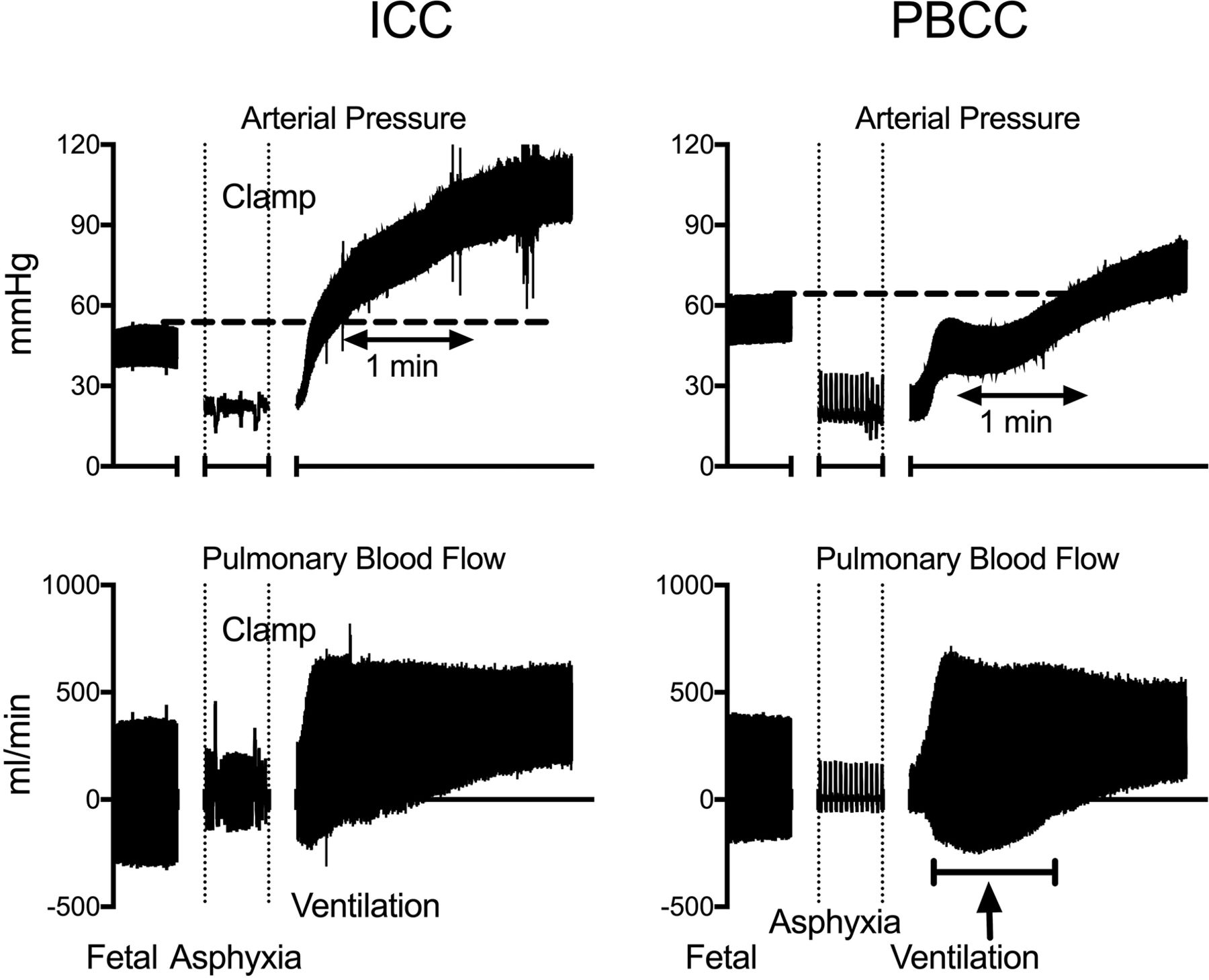
Real-time chart recording of carotid arterial blood pressure and pulmonary blood flow (PBF) from representative lambs undergoing ICC or PBCC. Note the significant overshoot in arterial blood pressure in the ICC lamb which is not present in the PBCC lamb. Also note the maintenance of negative end-diastolic PBF (arrow). ICC, immediate cord clamping; PBCC, physiologically based cord clamping.
The time taken for blood pressure to return to preasphyxia levels was significantly shorter in ICC compared with PBCC lambs (mean±SD; ICC: 83.3±32.7 s; PBCC: 127.1±33.5 s: P=0.037). Mean, systolic and diastolic carotid arterial pressures were significantly higher in ICC lambs than PBCC at 70 s after ventilation onset (figure 3). Mean and diastolic arterial blood pressures remained significantly elevated at 30 min in ICC lambs compared with PBCC lambs. From 140 s after ventilation onset, mean and systolic arterial blood pressures in ICC lambs were markedly higher (mean of 90–100 mm Hg) than the fetal values. This is indicative of a marked overshoot of blood pressure, with the average increase in mean arterial blood pressure being 39.3 mm Hg (range 30.7–44.2 mm Hg) greater than the respective fetal value. While PBCC lambs also displayed an overshoot in mean arterial pressure, the average overshoot was only 17.6 mm Hg (range 6.3–23.8 mm Hg), which was significantly lower (P<0.001) than that observed in ICC lambs.

(A–C) Mean, systolic and diastolic arterial pressure, (D–F) mean, peak systolic and end-diastolic PBF, and (G–I) mean, peak systolic and end-diastolic CaBF measured prior to delivery (fetal (F), at the end of the asphyxial period (A) and regularly in lambs undergoing ICC (open squares) or PBCC (closed circles)). *Indicates significant difference (P<0.05) between PBCC and ICC lambs at that time point. #Indicates trend (P<0.07). Values are mean±SEM. CaBF, carotid arterial blood flow; CBF, cerebral blood flow; ICC, immediate cord clamping; PBCC, physiologically based cord clamping; PBF, pulmonary blood flow.
Mean PBF was higher in ICC compared with PBCC lambs between 90 and 180 s after ventilation onset. The higher mean PBF occurred due to a significantly lower end-diastolic PBF in PBCC lambs between 1 and 10 min after ventilation onset (figure 3); peak systolic PBF was not different between groups. This indicates that PBCC lambs retain a significantly greater right-to-left shunt compared with ICC lambs, while the umbilical cord remained unclamped.
Mean, peak systolic and end-diastolic CaBF was not different between groups immediately after ventilation onset. However, mean, peak systolic and end-diastolic CaBF was significantly higher in PBCC lambs 20 and 30 min after ventilation onset compared with ICC lambs (figure 3). CaBF resistivity index was not different between groups, indicating no difference in downstream vascular resistance (data not shown).
Femoral arterial blood flow was significantly higher in ICC lambs between 1 and 3 min after delivery, but was not different thereafter. Peak systolic and end-diastolic femoral blood flow was not different between groups.
Cerebrovascular integrity and permeability
The number of blood vessels with protein extravasation in the subcortical and grey matter was significantly higher in ICC lambs compared with PBCC lambs (by 86% and 47%, respectively; figure 4). No difference was observed in the periventricular white matter.

Number of blood vessels with protein extravasation in the subcortical and periventricular white matter and the grey matter in asphyxiated lambs undergoing physiologically based cord clamping (PBCC) or immediate cord clamping (ICC). *Indicates significant difference from PBCC lambs. Values are mean±SEM.
We measured mRNA expression of key tight junction proteins critical for maintaining integrity of the blood–brain barrier. We observed decreases in iNOS, claudin-1 and claudin-5 in the subcortical and periventricular white matter and occludin within the subcortical white matter, but significance was only obtained for claudin-5 and occludin within the subcortical white matter (figure 5).

Relative mRNA expression of key tight junction proteins integral for vascular integrity within the subcortical white matter (A) and periventricular white matter (B) in asphyxiated lambs undergoing PBCC or ICC. *Indicates significant difference from PBCC lambs. Data are expressed relative to PBCC lambs. Values are mean±SEM. ICC, immediate cord clamping; iNOS, inducible nitric oxide synthase; PBCC, physiologically based cord clamping.
Discussion
The fetal response to asphyxia includes a redistribution of cardiac output to increase blood flow and preserve oxygen delivery to vital organs, particularly the brain. While lifesaving in utero, this cardiovascular adaptation could increase the risk of cerebrovascular injury immediately after birth when the cerebral vascular bed is maximally vasodilated.12 Here we investigated the effects of PBCC on the cardiovascular and cerebral physiology during resuscitation from asphyxia. We found that cardiac output and oxygenation were restored similarly between groups; however, PBCC prevented a marked overshoot in blood pressure after ventilation onset, and reduced cerebrovascular injury.
Aeration of the lung is the most critical component for survival of newborn infants at birth as it allows the onset of pulmonary gas exchange and triggers a rapid increase in PBF, which is required to sustain cardiac output following cord clamping.27 As such, the timing of ventilation onset relative to cord clamping is becoming increasingly recognised as an important component of newborn care, particularly in infants who are apnoeic at birth. ICC prior to lung aeration separates the infant from oxygen supply via the placental circulation, and deprives the left heart of venous return, causing a sudden loss in systemic cardiac output.17 The only way that cardiac output can be restored is by increasing PBF in response to lung aeration, which allows pulmonary venous return to take over as the source of left ventricular preload and hence left ventricular output.17
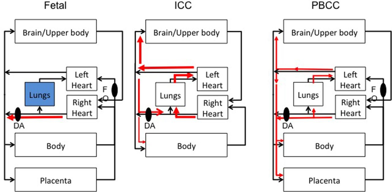
In our previous study, we found that a sustained inflation was very effective at restoring cardiovascular function in severely asphyxia newborn lambs, although the resulting rebound hypertension was thought to increase the risk of cerebral vascular leakage.28 In this study, we found that ventilating and resuscitating lambs while the umbilical cord remained open, greatly mitigated the rebound hypertension and reduced cerebral vascular leakage compared with ICC lambs. Indeed, mean arterial pressures were over 15 mm Hg (68.4±6.2 vs 51.4±1.9 mm Hg) higher in the ICC lambs compared with PBCC lambs at 2 min after ventilation onset. This was due to the presence of the highly compliant, low-resistance placental vascular bed remaining within the systemic circuit in PBCC lambs during the recovery in cardiovascular function. In this regard, the placenta acts as a pressure relief valve that competes with (1) PBF for right ventricular output and (2) with flow to the upper body for left ventricular output (figure 6). Consistent with this explanation, we found that the increase in PBF was much greater in ICC lambs than PBCC lambs. This is because the placenta provides a low-resistance alternative pathway for right ventricular output, allowing blood to bypass the pulmonary circulation and enter the descending aorta via the ductus arteriosus. Indeed, retrograde end-diastolic PBF continued until at least 5 min after ventilation onset in PBCC lambs, in contrast to ICC lambs, which is indicative of continued right-to-left flow across the ductus arteriosus. Femoral arterial blood flow was also higher in ICC in the first minutes after birth, again highlighting the competition for flow between the placenta and newborn circulation.

The fetal circulation is unique because the placenta provides blood flow for both right ventricular output (RVO) and left ventricular output (LVO), via the foramen ovale (FO) since the lungs are filled with liquid and the majority of RVO bypasses the lungs through the DA. Removal of the placenta with ICC and subsequent ventilation results in a rapid uncontrolled increase in pulmonary blood flow (PBF) resulting in a large increase in cardiac output and cerebral blood pressure. Ventilation before ICC (PBCC) allows for a controlled increase in PBF, LVO and subsequently reduced cerebral blood pressure as the placenta provides an alternative path for both RVO and LVO. DA, ductus arteriosus; ICC, immediate cord clamping; PBCC, physiologically based cord clamping.
Severe asphyxia, irrespective of whether it is induced by occlusion of the internal iliac artery or umbilical cord in near-term lambs, results in widespread pathologies to the brain. These include cell death, white matter disruption, oxidative stress, intraparenchymal haemorrhage and inflammation, which is consistent to that observed in infants with Hypoxic-Ischemic Encephalopathy.29–31 The resultant injury also delays the lamb’s abilities to (1) use all four legs, (2) attain a standing position, (3) find the udder, and (4) successfully suckle—compared with control lambs.29
It is very interesting that despite a >20 mm Hg difference in arterial pressure at 5 min following ventilation onset, CaBF was similar between groups. CaBF is controlled primarily at the level of the resistance vessels (arterioles), which also act in response to high blood pressures to limit flow into the brain and protect the more delicate downstream microvessels from high pressures and flows. However, this autoregulatory process is relatively slow (2–3 min), particularly in the immature cerebral vascular bed, making CaBF pressure passive in response to rapid changes in pressure.32 Thus, when the cerebral vascular bed is maximally vasodilated, large and rapid changes in pressure will directly impact on the small delicate cerebral microvessels. It is not surprising, therefore, that we demonstrated degradation of key blood–brain barrier proteins coupled with significant increases in cerebrovascular leakage in the subcortical and grey matter of the ICC brains compared with PBCC. Protein extravasation is indicative of a breakdown in the blood–brain barrier that is associated with increased risk of haemorrhage. It is possible that the similar CaBF in ICC lambs despite the higher arterial pressure was due to higher vasoconstriction in response to higher blood pressure. However, we do not believe this to be the case for a number of reasons. First, blood pressures were not different until 1 min after ventilation, but by 2 min there was a large difference in pressure and no difference in flow. This is too rapid for autoregulatory vasoconstriction and there is no evidence of this in values measured between the 1 and 2 min period. Second, if the ICC lambs had rapidly vasoconstricted in response to the higher pressure, this would have protected the cerebral microvessels from injury and as such we would have observed similar levels of vascular injury between the groups.
An alternative explanation for the finding of similar CaBFs is that the cerebral vascular bed was maximally dilated and that cerebral blood flow was at a maximum in both groups. Thus, despite a large difference in input pressure between the two groups, no difference in CaBF was measured. In this situation, vasodilated resistance vessels would likely expose the delicate microvasculature to elevated pressures causing damage, and the higher the pressures, the greater the damage. Interestingly, CaBF was lower at 20 and 30 min in ICC lambs compared with PBCC lambs, which coincided with a lower (of ~8%; P=0.3) SctO2 and higher cerebral oxygen extraction. We previously demonstrated in growth-restricted lambs that cerebrovascular injury resulted in reduced CaBF, increased protein extravasation and higher oxygen extraction.33 The similar findings in this study suggest that cerebrovascular injury results in increased vasoreactivity, ultimately reducing cerebral perfusion, as a protective effect to prevent increased injury. While the mechanism for this is unknown, it could be mediated by withdrawal of astrocyte end-feed from the blood vessel walls due to haemodynamic injury, resulting in cerebrovascular breakdown and increased leakage.34 However, more research is required to fully elucidate the cause of cerebrovascular dysfunction in this model.
It is pertinent that we have been able to show protein extravasation injury, an early marker of haemorrhage, in association with perturbations in cerebral blood pressure during resuscitation from asphyxia. Numerous studies in preterm infants have shown that fluctuations in cerebral blood flow, cerebral blood flow passivity or abnormal blood pressure are associated with cerebrovascular injury, particularly IVH (reviewed by Ballabh35). However, this study shows that a lack of cerebral autoregulation induced by asphyxia also predisposes the term newborn to cerebrovascular injury. While prevention of the primary asphyxial injury is the obvious target for intervention, it is also important that neonatal resuscitation minimises the consequences of this injury. Simple, but effective, interventions such as PBCC are particularly relevant for the developing world where the burden of asphyxia injury remains very high.
We have shown that it is feasible to initiate positive pressure ventilation while the asphyxiated newborn is still attached to the umbilical cord, while still allowing restoration in oxygenation and cardiac output at the same rate and in a similar fashion as lambs resuscitated following ICC. As such, we could find no benefit of ICC in these asphyxic lambs. Lambs in the PBCC group were ventilated for 10 min prior to cord clamping to determine the effect of having the low-resistance placental vascular bed in circuit during the recovery phase from asphyxia. We found that the rebound hypertension peaked between 5 and 10 min after ventilation onset, indicating that the benefits of PBCC in this subset of infants are likely to require an extended period (10 min or more) before cord clamping. Recent studies in babies at risk of resuscitation demonstrate the feasibility of resuscitation on the umbilical cord for >5 min (348±115 s), with improvements in blood pressure, cerebral oxygenation and reduced oxygen extraction compared with a 1 min cord clamping interval.36 More studies are required to determine the optimal duration of ventilation prior to umbilical cord clamping as well as the development of clinical indicators that can guide when cord clamping should occur.
This animal work has some limitations in transition to the clinical setting. Clinical extrapolation of this work is limited by anaesthesia, caesarean section delivery and the induction of asphyxia by reducing maternal placental blood flow. The underlying physiology will be conserved in the clinical setting but the conservation of placental blood flow during vaginal deliveries will require further investigation. In regard to clinical applicability, based on our findings, the primary issue of whether PBCC has any benefit over ICC in asphyxiated infants is whether or not flow can continue in the umbilical vessels during the recovery. If flow cannot occur, for instance, if there is a knot in the cord and it is occluded, then PBCC has no clear benefits and ICC is preferable. Other situations in which PBCC would not be advisable include placental haemorrhage as the infant would then be at risk of losing blood volume during PBCC. However, placental abruption or nuchal cords (as long as umbilical flows can be restored) are not necessarily an indication for ICC. This is because the placenta is not required for gas exchange as the lungs have taken over this role. Indeed, it is not uncommon for very preterm infants to be delivered with the placenta and fetal membranes still intact. Clearly, in this circumstance, the placenta will not be functioning in terms of gas exchange, but it will continue to serve as a highly compliant vascular bed that can mitigate the rebound hypertension and potential brain injury associated with the postasphyxia recovery.
Summary
In summary, ICC followed by ventilation resulted in significant postasphyxia rebound tachycardia and hypertension. Thus, ICC exposes the infant’s brain to higher pressures during the immediate postasphyxial period, thereby increasing the risk of cerebrovascular injury. PBCC restored cardiac output and oxygenation in an identical time frame as ICC, but protected the asphyxiated brain from cerebrovascular injury likely by mitigating the postasphyxia rebound hypertension in the setting of poor cerebral autoregulation. PBCC may be a more suitable option for resuscitation of asphyxiated newborns compared with ICC.
Acknowledgments
The authors would like to thank Jade Barbuto, Karyn Rodgers and Alison Moxham for their technical support.
References
Footnotes
Contributors Substantial contributions to the conception or design of the work, or the acquisition, analysis or interpretation of data: GRP, DAB, SKB, SLM, MK, AWG, ABP, SBH. Drafting the work or revising it critically for important intellectual content: GRP, DAB, SKB, SLM, VS, MK, AWG, DL, ABP, SBH. Final approval of the version published: GRP, DAB, SKB, SLM, VS, MK, AWG, DL, ABP, SBH. Agreement to be accountable for all aspects of the work in ensuring that questions related to the accuracy or integrity of any part of the work are appropriately investigated and resolved: GRP, DAB, SKB, SLM, VS, MK, AWG, DL, ABP, SBH.
Funding This research was supported by the National Institute of Health (R01HD072848-01A1), the Research Foundation of the Cerebral Palsy Alliance, a joint National Heart Foundation of Australia and National Health and Medical Research Council (NH&MRC) Research Fellowship (GRP: 1105526), an NH&MRC Research Fellowship (SKB: 545921), an NHMRC-Australian Research Council Dementia Research Development Fellowship (SKB: 1110040), Australian Research Council Future Fellowship (SLM) and the Victorian Government’s Operational Infrastructure Support Program.
Competing interests None declared.
Ethics approval Experimental procedures were approved by the Monash Medical Centre Animal Ethics Committee A, Monash University.
Provenance and peer review Not commissioned; externally peer reviewed.