Article Text

Abstract
Objective: To assess the time course of recovery of severely abnormal initial amplitude integrated electroencephalographic (aEEG) patterns (flat trace (FT), continuous low voltage (CLV), or burst suppression (BS)) in full term asphyxiated neonates, in relation to other neurophysiological and neuroimaging findings and neurodevelopmental outcome.
Methods: A total of 190 aEEGs of full term infants were reviewed. The neonates were admitted within 6 hours of birth to the neonatal intensive care unit because of perinatal asphyxia, and aEEG recording was started immediately. In all, 160 infants were included; 65 of these had an initial FT or CLV pattern and 25 an initial BS pattern. Neurodevelopmental outcome was assessed using a full neurological examination and the Griffiths’ mental developmental scale.
Results: In the FT/CLV group, the background pattern recovered to continuous normal voltage within 24 hours in six of the 65 infants (9%). All six infants survived the neonatal period; one had a severe disability, and five were normal at follow up. In the BS group, the background pattern improved to normal voltage in 12 of the 25 infants (48%) within 24 hours. Of these infants, one died, five survived with moderate to severe disability, two with mild disability, and four were normal. The patients who did not recover within 24 hours either died in the neonatal period or survived with a severe disability.
Conclusion: In this study there was a small group of infants who presented with a severely abnormal aEEG background pattern within six hours of birth, but who achieved recovery to a continuous normal background pattern within the first 24 hours. Sixty one percent of these infants survived without, or with a mild, disability.
- aEEG, amplitude integrated electroencephalography
- BS, burst suppression
- CLV, continuous low voltage
- CNV, continuous normal voltage
- DNV, discontinuous normal voltage
- FT, flat trace
- MRI, magnetic resonance imaging
- asphyxia
- amplitude integrated electroencephalography
- background pattern
- neurodevelopmental outcome
Statistics from Altmetric.com
- aEEG, amplitude integrated electroencephalography
- BS, burst suppression
- CLV, continuous low voltage
- CNV, continuous normal voltage
- DNV, discontinuous normal voltage
- FT, flat trace
- MRI, magnetic resonance imaging
During recent years the clinical use of amplitude integrated electroencephalography (aEEG) in the neonatal intensive care unit has increased. The aEEG background pattern correlates well with the standard EEG, and the method has been noted to have very good predictive value for neurodevelopmental outcome in term neonates after perinatal asphyxia, especially in the first hours after birth.1–,7 This early assessment has been used for selection of infants who might benefit from neuroprotective therapy.8,9 It is also used for informing the parents about the likely prognosis and provides additional information for use in the decision to continue or withdraw intensive care in a severely asphyxiated infant. It is known that the background pattern can change during the first 24–48 hours after birth, especially in infants with a burst suppression pattern.5,10,11 Spontaneous recovery in aEEG background patterns after perinatal asphyxia has not yet been described in detail.
The aim of this study was to assess the recovery of aEEG patterns in full term asphyxiated neonates who present with a severely abnormal initial aEEG (flat trace (FT), continuous low voltage (CLV), or a burst suppression (BS) pattern). We hypothesised that children in whom an abnormal background pattern rapidly recovered would have a chance of a normal neurodevelopmental outcome.
METHODS
Between 1992 and 2002, all full term neonates with perinatal asphyxia, who were admitted to the neonatal intensive care unit at Wilhelmina Children’s Hospital (a tertiary referral centre) had an aEEG recorded for at least 24 hours (range 24 hours to 7 days). Asphyxia was diagnosed if they met at least three of the following criteria:
signs of intrauterine asphyxia, as indicated by late decelerations on fetal monitoring or meconium stained liquor
arterial cord blood pH <7.10
delayed onset of spontaneous respiration
Apgar score of ⩽5 at five minutes
multiorgan failure.
For aEEG recording, the CFM 4640 (Lectromed, Letchworth, Hertfordshire, UK) was used. The CFM records a single channel EEG from bilateral parietal electrodes. The filtered signal is rectified, smoothed, and amplitude integrated before it is printed out at slow speed (6 cm/h) at the cot side.4,5 For pattern recognition, we used the following criteria4,5,12:
FT: very low voltage, mainly inactive (isoelectric) tracing with activity below 5 μV;
CLV: continuous background pattern of very low voltage (around or below 5 μV);
BS: discontinuous background pattern periods of very low voltage (inactivity) intermixed with bursts of higher amplitude;
discontinuous normal voltage (DNV): discontinuous trace, where the voltage is predominantly above 5 μV;
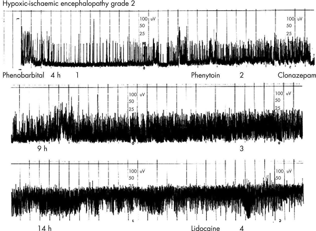
continuous normal voltage (CNV): continuous activity with voltage 10–25 (−50) μV (fig 1⇓).

Example of an amplitude integrated electroencephalographic pattern, which shows recovery within 24 hours. The different background patterns are: 1, flat trace; 2, burst suppression; 3, discontinuous normal voltage; 4, continuous normal voltage.
We retrospectively reviewed the medical records of 190 infants in whom the aEEG recording was started within the first 6 hours of birth. Infants with congenital malformations or chromosomal abnormalities were excluded, and one patient had to be excluded because he was lost to follow up. Thus 160 full term asphyxiated infants were eligible for this study. The aEEG tracings were reviewed by two clinicians who were blinded to the outcome. The tracings were analysed for the background pattern during the first 6 hours after birth. If a depressed background pattern (BS/CLV/FT) was present, we followed the recovery. First we noted the time of recovery to a DNV pattern, as this pattern tends to be associated with a normal neurodevelopmental outcome.5 We subsequently noted the time of recovery to a CNV pattern. Administration of anti-epileptic drugs was also noted. Seventy neonates had a normal aEEG (CNV/DNV) pattern at the onset (fig 2⇓). For this study, we focused on a group of 65 neonates who had an initial FT or CLV pattern, and 25 neonates with an initial BS pattern within 6 hours of birth. The mean gestational age of the FT/CLV group was 40 weeks (range 37–42) and mean birth weight 3440 g (range 2170–4885). In the BS group, the mean gestational age was also 40 weeks (range 37–42), and the mean birth weight 3480 (range 2580–5140). All had a moderate to severe encephalopathy, grade 2 and 3 according to Sarnat and Sarnat.13 None of the patients were involved in an early intervention study.
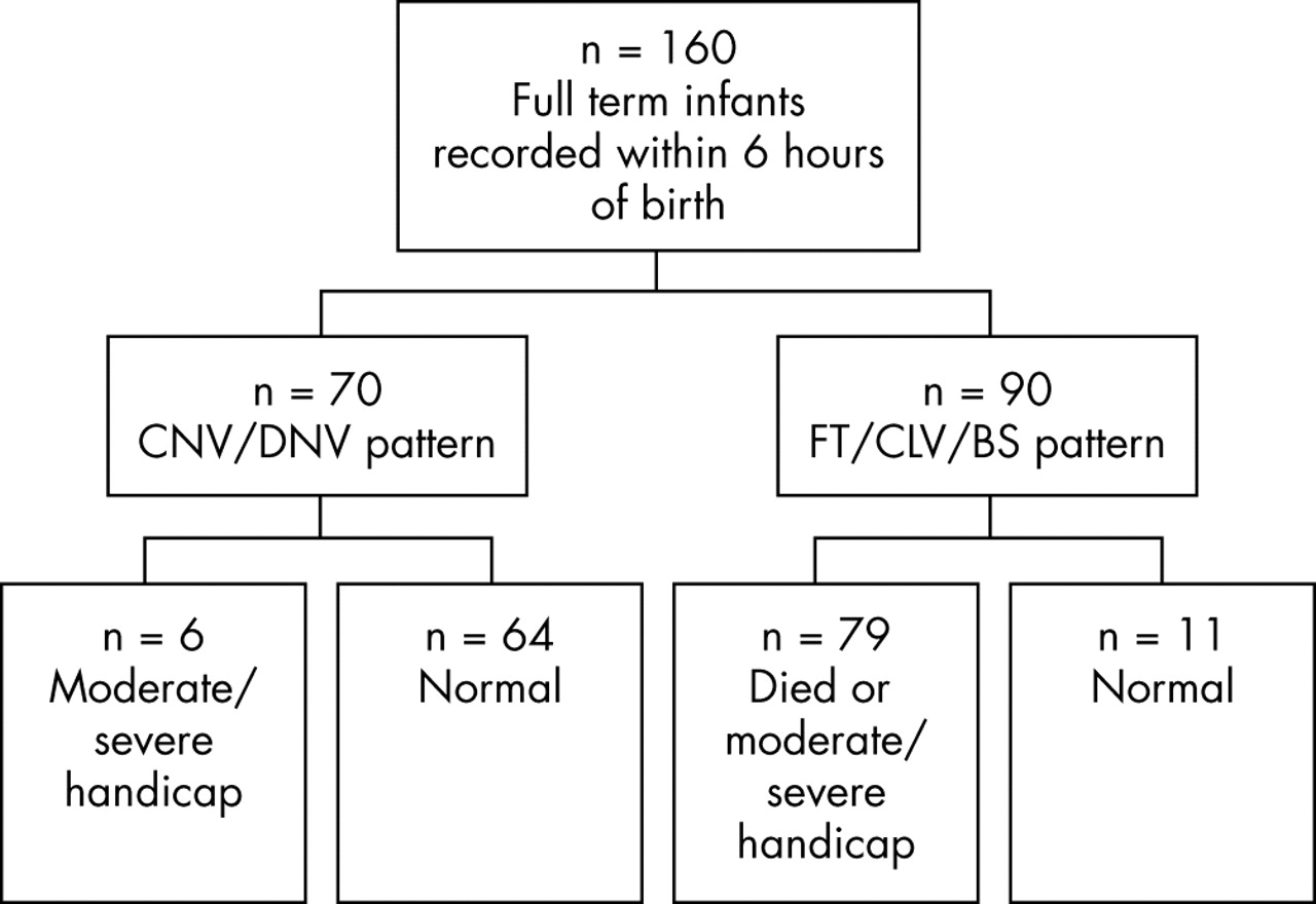
Flow diagram of 160 study patients. BS, Burst suppression; CLV, continuous low voltage; CNV, continuous normal voltage; DNV, discontinuous normal voltage; FT, flat trace.
All survivors were seen in our outpatient clinic. Neurodevelopmental outcome was assessed using the Griffiths mental developmental scale at postnatal ages of at least 24 months.14 The maximum age at follow up was 10 years. Neuromotor assessment was performed by a paediatric physiotherapist, who was blinded to the aEEG and other neonatal data.
Cerebral palsy was classified according to the criteria of Hagberg et al.15 Adverse outcome was defined as death in the neonatal period, cerebral palsy, or a developmental quotient <85 on the Griffiths scale at 2 years of age. Maximal locomotor function of those with cerebral palsy was graded according to a simplified version of the classification suggested by Palisano et al.16 At 2 years, a distinction was made between: (a) walking independently without restrictions—can take more than 10 steps without any help; (b) sits independently—infant maintains floor sitting and may pull to stand and take steps holding on to furniture; (c) cannot sit—is unable to maintain anti-gravity head and trunk control in prone and sitting positions.
Most of the infants had a magnetic resonance imaging (MRI) scan during the first 2 weeks of birth (median age 9 days, range 5–15; up until 1999 only conventional images; since 1999 diffusion weighted images also). Abnormal findings in cortex, basal ganglia, thalamus, white matter, and posterior limb of the internal capsule seen after a hypoxic-ischaemic insult were assessed.17–,19
A statistical analysis was performed using SPSS for windows version 11.5. For group comparison, the Mann-Whitney U test was used for continuous variables and Fisher exact or χ2 tests for categorical variables. Sensitivity, specificity, and positive and negative predictive values of the test were calculated. p<0.05 was regarded as significant.
RESULTS
Figure 2⇑ shows a flow diagram of the 160 study patients. Of the 70 patients with an initial normal or slightly abnormal (CNV/DNV) pattern within the first 6 hours of birth, all survived. Six had a moderate or severe handicap, and 64 were normal at follow up. In the group of 90 infants with an abnormal background pattern, only 11 had a normal outcome (p<0.001, χ2 test).
We calculated the sensitivity, specificity, and positive and negative predictive values for FT/CLV together with BS at the age of 6 hours for a poor neurodevelopmental outcome. Table 1⇓ shows the results, together with comparative results of previous studies, also calculated 6 hours after birth. Thirty eight patients in the study of Toet et al5 were also included in this study.
Predictive values of burst suppression together with flat trace/continuous low voltage for poor outcome in studies carried out within 6 hours of birth
The positive predictive value for a normal outcome was 91% for the infants with a CNV pattern within 6 hours of birth, compared with 61% in infants who showed recovery to a CNV pattern within the first 24 hours of birth.
FT/CLV group
A total of 65 infants had a FT or CLV background pattern when aEEG was started within the first 6 hours of birth. In six (9%), the pattern improved to CNV within 24 hours (fig 3⇓).

Flow diagram of patients who had a flat trace (FT) or continuous low voltage (CLV) background pattern within the first 6 hours of birth.
Table 2⇓ shows the problems during delivery, Apgar scores, and aEEG data. All received one or more anti-epileptic drugs during the first 24 hours. All received phenobarbital as the first drug. In three infants (2, 5, and 6), phenobarbital was given as prophylaxis after birth asphyxia, but all these infants developed seizures later on. The infants with a normal outcome received one or two anti-epileptic drugs, in contrast with the infant with a poor outcome, who received four different anti-epileptic drugs. The 59 infants without recovery during the first 24 hours, all died during the first week of life from severe hypoxic-ischaemic encephalopathy, assessed in more detail using standard EEG, evoked potentials, ultrasonography, and MRI. In 22 infants (38%), postmortem examination was also performed, and the presence of extensive hypoxic-severe ischaemic damage was seen in all.
Amplitude integrated electroencephalography data and outcome of infants with recovery of flat trace/continuous low voltage pattern
Neurodevelopmental outcome
Of the six survivors, one infant showed severe learning disabilities, quadriplegia, and epilepsy at follow up. Five infants showed a normal neurodevelopmental outcome at a minimum age of 2 years. Of these infants, two were seen back in our outpatient clinic after these 2 years, one at 5 and one at 6 years of age. Both showed normal development.
Table 3⇓ shows the results of the other diagnostic techniques used in these infants. In the infant who survived with a severe disability, the ultrasound and the MRI showed severe abnormalities in the central grey nuclei. In the infants who were normal at follow up, additional neurophysiology and neuroimaging techniques showed mild abnormalities.
Results of other examinations in the group with flat trace/continuous low voltage patterns
BS group
A total of 25 infants had an initial BS background pattern. In this group, the pattern improved to CNV in 10 (40%) of the infants within 24 hours and in two within 48 hours, but the latter two had shown recovery to a DNV pattern within 24 hours (fig 4⇓).

Flow diagram of patients who had a burst suppression (BS) background pattern within the first 6 hours of birth.
Similar to the other group, all infants received at least one anti-epileptic drug during the first 24 hours, and in three infants (1, 8, and 9) phenobarbital was given as prophylaxis. In this group, the difference in the number of anti-epileptic drugs used in relation to outcome was not so clear. Most of the infants with an unfavourable outcome received three or more anti-epileptic drugs. Table 4⇓ shows delivery and aEEG data of this group.
Amplitude integrated electroencephalography data and outcome of infants with recovery of burst suppression pattern
In one infant, the background pattern initially recovered to CNV at 21 hours after birth, but 38 hours after birth an acute deterioration in the clinical condition was seen. The aEEG deteriorated to a BS pattern, and the infant subsequently died.
Neurodevelopmental outcome
Of the 11 survivors, four showed a normal development: one at the age of 2, two at the age of 3, and one at the age of 5 years. Two infants had a mild disability; both had a normal developmental quotient at 3 years but they had problems with language and speech development. Five infants had a severe handicap: one had ataxic cerebral palsy and epilepsy with a developmental quotient of 85 at the age of 2 years, and the others had dyskinetic cerebral palsy at the age of 2 years. Of the 13 infants with a persistent BS pattern after 24 hours, 10 died, and three survived with a severe disability. They all showed further deterioration of the background pattern to CLV or FT, and six of them had persistent seizures despite extensive anti-epileptic medication.
Table 5⇓ shows the results of the other examinations. The patient who died did not have an MRI because he was too ill to be transported to the MRI unit. In the infants with a handicap, ultrasound and MRI showed moderate to severe abnormalities. In the infants who were normal at follow up, there were also abnormalities, but most were mild.
Results of other examinations in the burst suppression group
Comparison of infants with FT/CLV and those with a BS pattern
In the FT group, five of the six (83%) infants with recovery had a normal outcome. In the BS group, six of the 12 (50%) infants with recovery had a normal or slightly abnormal outcome (p = 0.600, Fisher’s exact test).
The median time interval from birth to onset of DNV pattern in infants with a FT/CLV pattern was 6.5 hours, and in the BS group it was 9 hours (p = 0.637, Mann-Whitney U test). For the onset of the CNV pattern, the median time interval was 11 hours in the FT/CLV group and 11.5 hours in the BS group (p = 0.604, Mann Whitney U test).
Looking at the infants with an initially poor background pattern, those who showed recovery had a better outcome than those who had a persistently abnormal background pattern (p<0.001, Fisher’s exact test). We also found this in the FT/CLV group and BS separately (p<0.001 and p<0.002 respectively, Fisher’s exact test).
For infants with an abnormal aEEG within the first 6 hours of birth, lack of recovery within 24 hours, combined with abnormalities on a standard EEG, evoked potentials, ultrasonography, and MRI, predicted neonatal death or poor outcome in 59/65 infants in the FT/CLV group compared with 13/25 in the group with the BS background pattern (p<0.001, χ2 test).
DISCUSSION
In this study, we describe the recovery of an initially severely abnormal aEEG background pattern in 18 out of 90 full term infants with a diagnosis of perinatal asphyxia, in whom aEEG recording was started within 6 hours of birth. Early recovery of the background pattern was observed in only six out of 65 (9%) infants who presented with a FT/CLV background pattern and in 12 of 25 (48%) infants with a BS background pattern. Eleven of these 18 (61%) infants with a rapid recovery of the background pattern had a normal or mildly abnormal outcome (nine had a normal outcome and two a mildly abnormal outcome). Five of these 11 infants had a FT/CLV background pattern. Although rapid recovery of the background pattern was more common in the BS group, this was less often associated with a normal outcome. This difference in outcome for these two background pattern groups is of interest. The insult around the time of delivery in the FT/CLV group was almost invariably acute and severe, but presumably of shorter duration than in those with a persistent FT/CLV background pattern and in those with a BS pattern. As six of the 12 infants in the BS group had an abnormal outcome in spite of rapid recovery, aEEG by itself was insufficient to accurately predict neurodevelopmental outcome. Other modalities such as ultrasonography, MRI, and evoked potentials were required to provide a more accurate prediction. In the infants who survived and developed dyskinetic cerebral palsy, abnormalities of the basal ganglia and thalami were seen on MRI, suggestive of acute near total asphyxia as the underlying disorder.17,20–,23 MRI is, however, not a bedside technique, and abnormalities of the basal ganglia are not always visible during the first few days after birth, even with the use of diffusion weighted imaging.22,24,25 Evoked potentials showed delayed or absent responses in most of the infants who survived with moderate to severe disabilities. This is in agreement with previous studies, showing that evoked potentials are highly predictive.10,26,27 In two infants, the evoked potentials were normal, but MRI showed abnormalities. This would suggest that the lesions seen on MRI were of antenatal origin.
Although data on an initially inactive or severely abnormal EEG and a normal outcome do exist, they are scarce. Pezzani et al28 showed that recovery of an inactive EEG within the first 10 hours of birth does occur and can be associated with a normal outcome. Selton and Andre29 reported on one infant with an initially severely abnormal EEG 9 hours after birth, who recovered within 24 hours and had a normal outcome. Pressler et al30 reported on two infants with an inactive EEG during the first 8 hours after birth, who showed recovery within 12 hours of birth, and both infants had a normal outcome. Our data are in agreement with these studies and stress the importance of continuous monitoring beyond the first 12 hours of birth to assess whether rapid recovery occurs, even though this is not a common finding when a FT/CLV background pattern is seen.
We previously reported that the presence of a poor background pattern at 6 hours of age had a positive predictive value of 84% and negative predictive value of 91% for later neurodevelopmental outcome.5 In this larger group of infants, these data still hold true, with a positive predictive value of 88% and a negative predictive value of 91%. Another study, considering much smaller groups of patients, reported similar predictive values. Hellström-Westas et al4 studied 47 term asphyxiated infants who had aEEG monitoring started before 6 hours of life. BS, CLV, and FT were regarded as abnormal background patterns and predictive of poor outcome. They found a positive predictive value of 86% and negative predictive value of 96%.
All our infants received at least one, but more often at least two, anti-epileptic drugs during the first 24 hours. Phenobarbital was given as the first choice drug in all infants. In spite of administration of anti-epileptic medication, recovery of the aEEG pattern was noted. It has been reported that anti-epileptic drugs can lead to transient deterioration of the aEEG background pattern, and that this deterioration also depends on the severity of the preceding insult to the brain.4,5,31–,33 The fast recovery seen in this study therefore implies that the brain injury of some of our infants was not as severe as suggested by their initial aEEG findings. The infants in the FT/CLV group, with a normal outcome, received one or two anti-epileptic drugs, and those with a poor outcome received four different drugs. In the BS group, this difference was not so clear. It is known that seizures (clinical and electrographic) are often associated with an abnormal outcome, especially when combined with an abnormal background pattern.23,34,35
The aEEG in full term infants, during the first hours after perinatal asphyxia, can predict neurodevelopmental outcome, making it a very useful technique for early selection of infants for neuroprotective treatments. We have shown that there is a small group of infants who have a severely abnormal background pattern during the first 6 hours after birth, but recover to a normal background pattern within 24 hours. Almost half of the infants with a rapid recovery survived without or with a mild disability. Assessment of outcome took place at 24 months of age. As recently reported by Barnett et al,36 long term follow up of these children is of importance, as a normal score in the early years cannot preclude later neurological, perceptual-motor, or cognitive abnormalities.
Thus early recovery of an initially abnormal aEEG background pattern is of predictive value. However, in the case of a rapid recovery of an initially poor aEEG pattern, additional techniques are needed to make a further distinction between those with a good or a poor neurodevelopmental outcome. We would like to stress the importance of early monitoring, as there is a difference in predictive value of an initially normal background pattern compared with an initially abnormal pattern showing recovery within 24 hours of birth.
We recommend continuation of aEEG recording and intensive care treatment in the severely asphyxiated infant for at least 24 hours, as there is a chance of recovery with a good neurodevelopmental outcome. Long term follow up of these children is important.
Acknowledgments
LvR was supported by the Dutch Epilepsy Foundation. DO was supported by the Slovenian Ministry of Education, Science and Sport and a Huygens grant.
REFERENCES
Footnotes
Competing interests: none declared
Linked Articles
- Fantoms