Article Text

Abstract
Background Gastroschisis is associated with low birthweight, which is thought to be due to protein leakage into the amniotic fluid from herniated bowel, bowel inflammation, placental chorangiosis and villous oedema. Little is known about the growth trajectories of fetuses affected with gastroschisis compared to those of normal fetuses.
Methods A retrospective review of fetal ultrasound measurements (biparietal diameter (BPD), head circumference (HC), abdominal circumference (AC), and femur length (FL), n = 830 measurements, mean of 6 measurements per fetus) from 130 fetuses with gastroschisis at UCLH (1992–2012). Size data were compared with BMUS reference centiles and were expressed as a function of gestational age. Growth velocity was deduced by the change in z score for each measurement between adjacent visits.
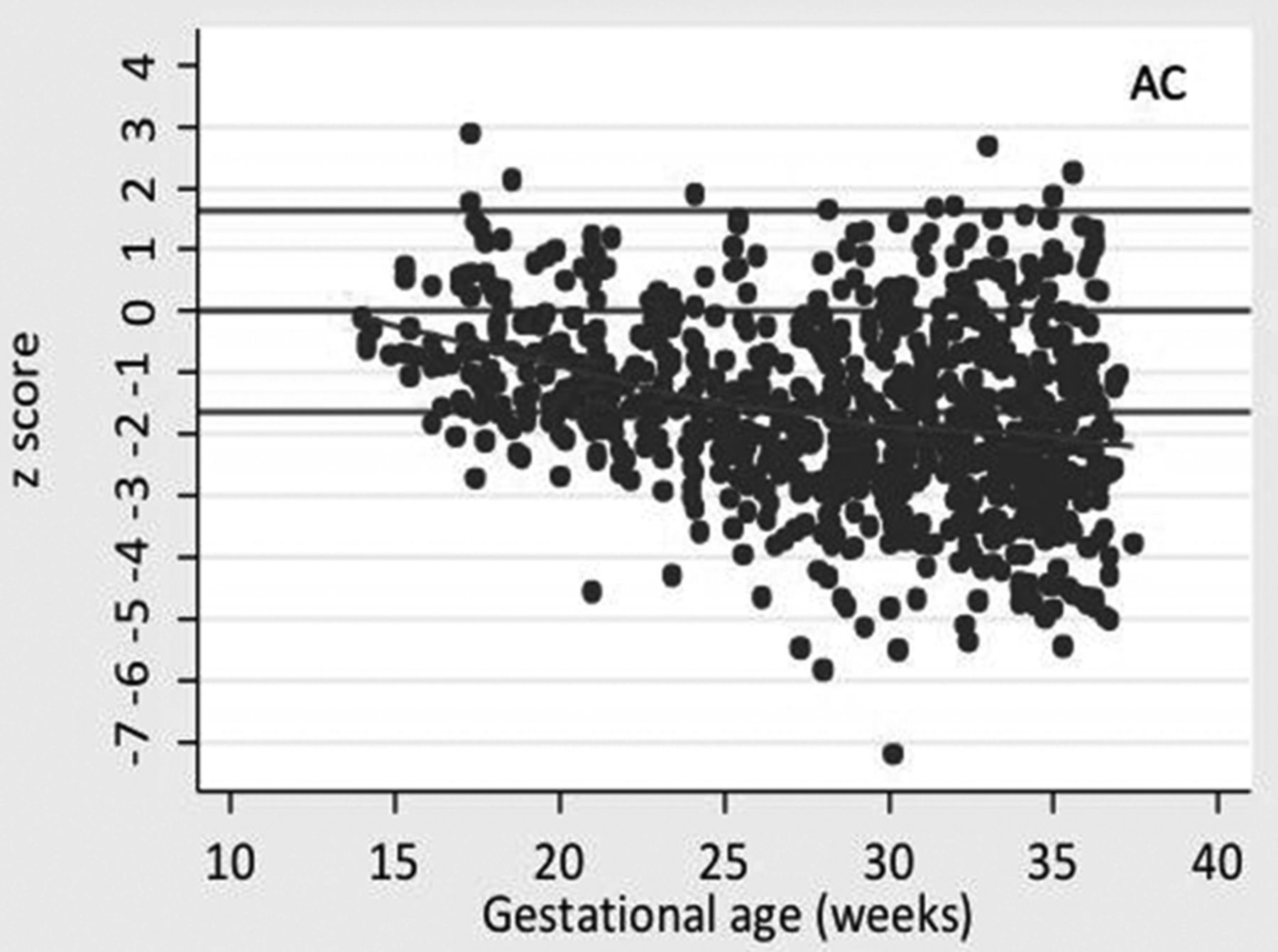
Results Average z scores fell from close to zero at early gestations (i.e. on average the same as normal fetuses) to -1 for BPD, HC, and FL and -2 for AC (Figure 1, AC as an example). For growth velocity, lowess curves for BPD, HC and AC were negative before 25 weeks suggesting that velocity is less than expected. After 25 weeks the mean was around zero suggesting that on average fetuses stay on the same centile (Figure 2, HC as an example). This pattern is not seen for FL where the lowess curve remains around zero for all gestation.

Average z score as a function of gestational age (AC)

Velocity (change in z score for each measurement against the GA at the point midway between the 2 measurements
Conclusions Fetuses with gastroschisis have a slower increase in BPD, HC and AC measurement before 25 weeks compared with normally formed fetuses but after this time velocity normalises.